


With severe internal diseases, poor nutrition, as well as with age, nail growth slows down, its structure changes. Only a doctor can accurately determine the cause of the violation based on the results of tests and microscopic examination.
But to get an idea of what happens to the nails on the feet or hands, you can use photos with fungal diseases of various kinds.
Causes of nail deformity
Fungi, yeast -like fungi and dermatophyte fungi cause infectious nail disease (onychomycosis), which shows similar symptoms.
All types of toenail or fingernail fungus damage the nail plate, change its transparency, shine, color, this variety can be seen in the photos presented.
Changes in the nails occur not only with onychomycosis, but also with injuries, chronic paronychia (inflammation of the nail folds), psoriasis, hand eczema, dermatitis. Before concluding that there is a fungal infection, you need to consider all possible options.
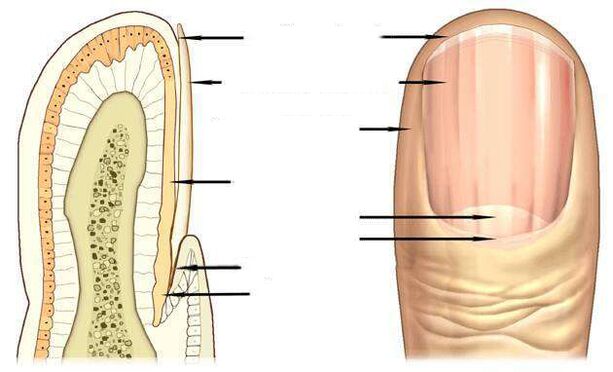
Signs of a fungal infection
The most informative signs of a fungal infection are discoloration of the nail plate, the presence of nail discharge, superficial changes - transverse, longitudinal grooves on the nail plate, point emphasis, thickening, destruction of the nail.
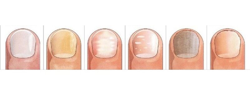
The pink color of a healthy nail is determined by the transparency of the nail plate and the blood vessels that appear through it. With onychomycosis, the nail loses its transparency, its color becomes brownish, yellow, more rarely green, black.
Candida fungi and dermatophytes cause onycholysis - the separation of the affected part of the nail. When infected with dermatophytes, onycholysis is observed from the edges of the isolated nail, and when infected with Candida, the nail is left behind the nail bed at the base, in the crescent area.
Symptoms of candidate fungus can be inflammation of the lateral periungual ridge - paronychia. The disease has a bacterial form caused by streptococci and staphylococci, as well as non -infectious - eczema, psoriasis, systemic vasculitis.
When the toenails are exposed to the fungus Trichophyton rubrum, the plate will be affected, as you can see in the photo, the roller will not be infected. The plate becomes yellowish, thickens strongly, the accumulated fungal mass is well distinguished beneath it.
Nail fungus due to dermatophyte infection
In 95% of all cases of nail fungus, the disease is caused by the dermatophytes Trichophyton rubrum and Trichophyton mentagrophytes.
Trichophyton rubrum infection
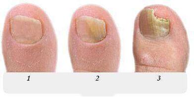
Onychomycosis begins when the fungus penetrates under the nail plate from the free edge. Fungal infections are indicated by the appearance of yellowish spots, uneven surfaces and collapses on the edges of distant (isolated) nails in the spot area.
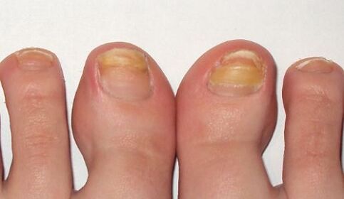
distal-lateral formTrichophyton rubrum dermatophyte fungal infection is common. In the photo, you can see that the stain caused by the introduction of the fungus is located along the lateral periungual nail folds.
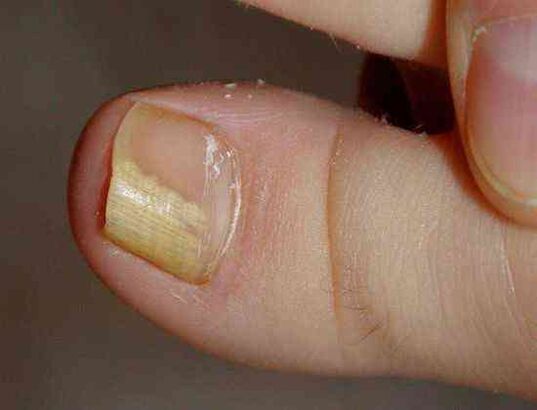
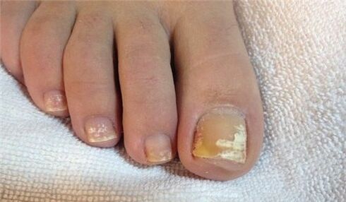
The fungus Trichophyton rubrum, as a rule, affects the big toes, causing hyperkeratosis - the accumulation of fungus between the nail plate and the nail bed, which looks like a loose yellowish mass in the photo.
At this stage, the fungus occupies an insignificant part of the nail, as in the photo presented, and with the help of local treatment it is possible to overcome the newborn onychomycosis.
Without treatment, the stain grows, gradually affecting the entire edge of the nail, and then moves to half a month. In the photo, the nail fungus looks like yellowish lines leading to the nail plate growth zone.
Withthe distal form of nail fungus, which is often found on big toes, yellowish spots of infection appear on the distal edge of the nail, in the middle, as seen in the photo.
In advanced stages of the fungus on the feet, some nails are affected, as in the photo, and treatment is no longer limited to local medications and pills. In addition to antifungal medications, nails undergo hardware cleaning, to remove the nail plate in whole or in part.
Long -term therapy with the use of all known antifungal agents and treatments should be performed on the feet, caused by Trichophyton rubrum, with hyperkeritosis, as can be seen in the photo.
Fungal infections with total damage to the nail spread to the entire area of the nail plate, the nail is completely destroyed.
Infection with another representative of the dermatophyte, the fungus Trichophyton mentagrophytes, can also cause nail infections on the entire nail.
Infection of Trichophyton mentagrophytes
With the overall defeat of the toenail with the fungus Trichophyton mentagrophytes, the nail plate is deformed, the photo shows that it thickens, changes its structure, collapses, yellowish spots appear all over its surface.
Infection of the nail with this dermatophyte usually causes superficial white onychomycosis on the big toes, more rarely on the little toes.
This fungus practically does not occur on fingernails, often causes interdigital dermatophytosis on the feet, as in the photo, and requires simultaneous treatment on the skin of the feet and nails.
Symptoms of a nail fungus infection, usually on the feet, are white spots of various sizes, as in the photo, which are reminiscent of leukonychia - a disease on the nail plate itself.
But unlike leukonychia, where white spots are caused by the appearance of air bubbles in the nail layer, white spots on fungal infections are the result of the activity of Trichophyton mentagrophytes.
Rarely, superficial white onychomycosis is caused by mold; in AIDS, the causative agent of this type of fungus can be Trichophyton rubrum and affects the nails on both feet and hands.
Changes in nails due to Candida infection
Fungus usually occurs in women, it attacks the nails on working hands, which are more often in contact with water.
For candidal onychomycosis, a proximal form of infection is characteristic, in which the fungus first affects the nail folds of the nail base, then penetrates into the growth zone and the base of the nail. Then gradually move along the nail from the base to the edges, capturing the growing area of the nail plate.
The causative agent of the disease in candidal onychomycosis is Candida albicans. The fungus attacks toenails and toenails, spreading from the half -moon zone at the base of the nail plate, to the free edges, as can be seen in the photo.
Signs of Candida nail infectionalbicans is inflammation of the nail folds (paronychia), separation of the cuticle from the nail plate, pain, discharge of pus when a bacterial infection is attached.
Candida albicans can penetrate nails and from their free edges. In this case, they discuss a form of distal infection, which is combined, as a rule, with cutaneous candidiasis.
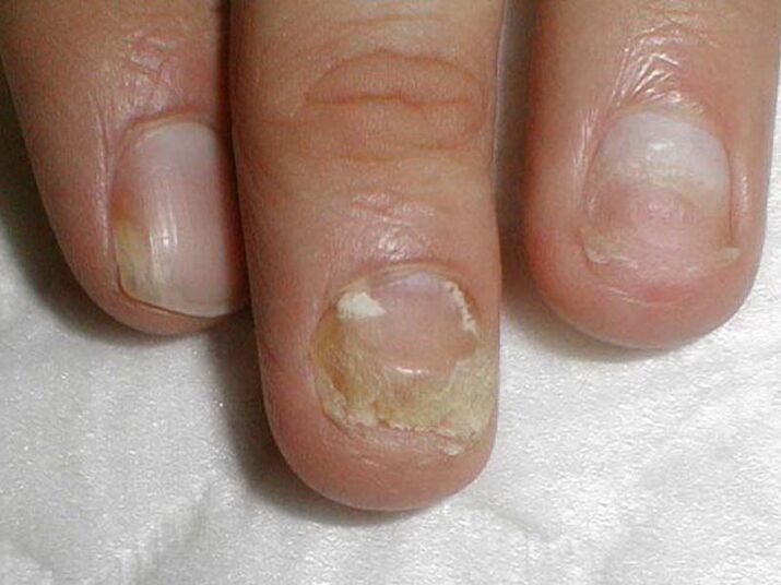
Treatment of candida fungus on fingernails and toenails with damage to more than half of the nail plate area, as in the photo, not only combats onychomycosis, but also measures to reduce candida activity in its natural reservoir - intestine, oral cavity, genital mucosa. . .
Invasion by mold
Fungi cause fungi more rarely than Candida or dermatophytes. The main symptom of a fungal nail infection is, as you can see in the photo, withchanging the color of the nail plate to blue, black, greenish.
Signs of toenail prints can be dark spots, spots on the nail plate, or, as in the photo, black longitudinal lines.
Preparations against fungus
Antifungal agents with fluconazole, ketoconazole, terbinafine, itraconazole, griseofulvin are used to treat nail fungus caused by dermatophytes, as in this photo.
Antifungal agents with terbinafine are effective for dermatophyte infections.
Antifungal agents with voriconazole are very active against dermatophytes.
Theseare used andto treat nail printon the feet, hands, andagainst candida yeast. The spectrum of action includes molds such as Aspergillum, Fusarium, Penicillium.
Itraconazole -based preparations overcome mold.
Fungal nail diseases
Gray colorsometimes appears on nailswith eczema. In this case, the nail plate can move away from the nail bed, which is observed with fungus.
Externally very similar tomanifestations of psoriasis onychomycosis. With this disease, not onlydiscoloration, but alsothickened nail plates.
Spot pressure is present on its surface, separation of the nail plate from the base of the nail is observed. But there is a difference from fungus: in psoriasis, the loose and healthy parts of the toenail are separated by pink and yellowing stripes over time.
The bluish colorgot nailswith pseudomonas nail infection. Frequent mechanical rubbing of the nail folds causes the appearance of shallow grooves, restlessness of the nail.
The white spots of leukonychia, whose appearance isassociated with metabolic disorders, can also be mistaken for a superficial white fungus with a large area.
Changes in color, shape of nails cause injury. Big toes are at great risk. Injured nails, also with fungus, thicken and darken.

The difference between injury and fungus is that changes during injury are observed only on the injured finger, the nail on the other finger is unchanged, do not be infected from a sore finger, as in onychomycosis.
The consequence of trauma can be the partial separation of the nail from the base of the nail, the formation of cavities, which, in unfavorable conditions, are quickly colonized by fungi.
Nail plates can be separated from the base of the nail under the influence of light (photoonicholysis), with iron deficiency anemia, hormonal diseases. Rupture, nail loss occurs with lichen erythematosus, bulosa dermatosis, nail trauma.
But you can finally make sure that the conclusion is correct and start treatment, you can only after getting help from a dermatologist - a dermatologist, or a mycologist - a doctor who treats fungal diseases.